


 收藏产品
收藏产品 产品信息
- Genetically modified cell lines best reflect MOA (Mechanism of Action)
- Higher activity and larger assay window for robust and reproducible cell-based bioassay
- Comprehensive application data to support assay development and validation
- Full tracible record, stringent quality control and validated cell passage stability
- Parental cell line legally obtained from internationally recognized cell resource bank and commercially licensed
- Global commercial license assistance whenever regulatory filing is required
描述(Description)
The HEK293/Human TL1A Stable Cell Line was engineered to express the full length human TL1A (Gene ID:9966). Surface expression of human TL1A was confirmed by flow cytometry.
应用说明(Application)
• Useful for cell-based TL1A binding assay
生长特性(Growth Properties)
Adherent
筛选标记(Selection Marker)
Puromycin (2 μg/mL)
培养基(Complete Growth Medium)
DMEM + 10% FBS
冻存液(Freeze Medium)
Serum-free cell cryopreservation medium
装量(Quantity)
1 vial contains at least 5×10^6 cells in 1 mL serum-free cryopreservation medium
存储(Storage)
Frozen in liquid nitrogen.
支原体检测(Mycoplasma Testing)
Negative
无菌检测(Sterility Testing)
Negative
使用说明(Instructions for Use)
See data sheet for detailed culturing and assay protocol.
产品数据图
Receptor Assay
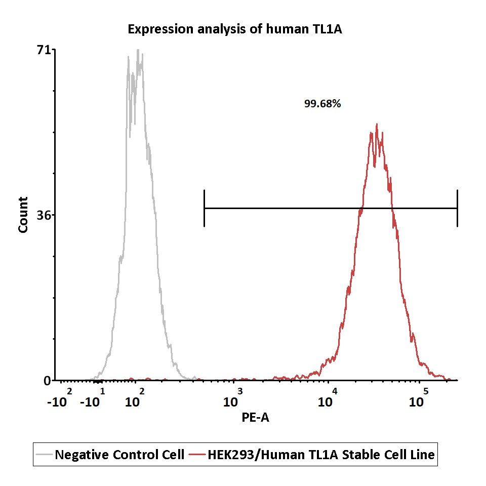
Expression analysis of human TL1A on HEK293/Human TL1A Stable Cell Line by FACS.
Cell surface staining was performed on HEK293/Human TL1A Stable Cell Line or negative control cell using anti-human TL1A Antibody followed by staining with PE anti-human IgG Fc Antibody.
Protocol
Passage Stability
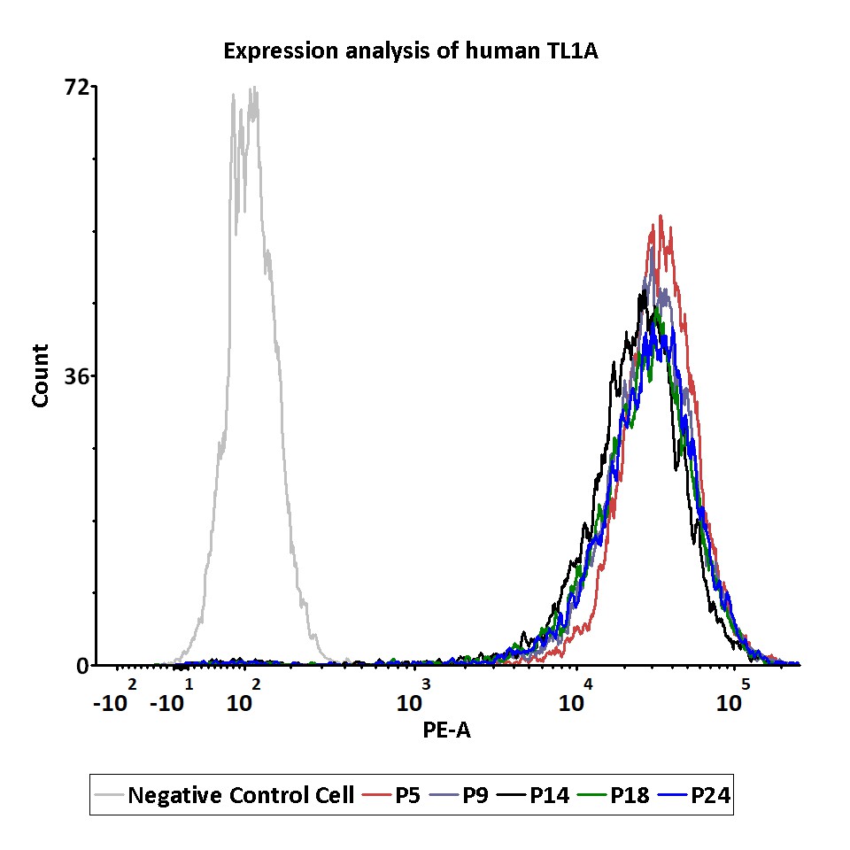
Passage stability analysis of human TL1A expression by FACS.
Flow cytometry surface staining of human TL1A on HEK293/Human TL1A Stable Cell Line demonstrates consistent mean fluorescent intensity across passage 5-24.
Protocol
产品评论 发表评论

背景
TNF-like cytokine 1A (TL1A) and its receptors, death receptor 3 (DR3) and decoy receptor 3 (DcR3) are members of the TNF and TNF receptor superfamilies of proteins, respectively. Binding of APC-derived TL1A to lymphocytic DR3 provides co-stimulatory signals for activated lymphocytes. DR3 signaling affects not only the proliferative activity of and cytokine production by effector lymphocytes, but also critically influences the development and suppressive function of regulatory T-cells. Whereas, DcR3 restricts the function of the TL1A/DR3 complex: attenuating T-cell activation and downregulating the secretion of pro-inflammatory cytokines. Together with DR3 and DcR3, TL1A constitutes a cytokine system that actively interferes with the regulation of immune responses.
 靶点信息
靶点信息  数据表和文档
  联系我们
联系我们 


 项目合作
项目合作