积分兑换
积分兑换 关注公众号
关注公众号
 收藏产品
收藏产品 产品详情
组分(Materials Provided)
Box A(2-8°C)IDComponentsSizeRIPO-BWM001K-C01Basal medium A22.5 mLRIPO-BWM001K-C02Basal medium B22.5 mLRIPO-BWM001K-C03Basal medium C90 mLBox B(-20°C)IDComponentsSizeRIPO-BWM001K-1-C01Supplement A-12 mLRIPO-BWM001K-1-C02Supplement A-20.5 mLRIPO-BWM001K-1-C03Supplement B-12 mLRIPO-BWM001K-1-C04Supplement B-20.5 mLRIPO-BWM001K-1-C05Supplement C-18 mLRIPO-BWM001K-1-C06Supplement C-22 mL产品描述(Product Details)
Human iPSC-Derived Cerebral Organoid Differentiation Kit (Ca. No. RIPO-BWM001K) allows hESC or hiPSC to differentiate into cerebral organoids. Cerebral organoids are three-dimensional in vitro models with a cellular composition and structural organization that is representative to the human cerebral regions. This kit can produce 96 cerebral organoids in three steps. Differentiation was carried out by forming EBs from PSC at an ultra-low attachment U shape 96 well plate, and then changing the medium according to the instructions. Organoids generated using Human iPSC-Derived Cerebral Organoid Differentiation Kit (Ca. No. RIPO-BWM001K) feature various types of neurons (including TH postive neurons) and glia cells (including OLIG2 and IBA1 positive cells).
产品特征(Product Specification)
The basic medium of this kit is a serum-free, well-defined medium with minimal batch variation to which differentiation factors are added. This medium does not contain antibiotics, the addition of which may affect organoid differentiation.
存储(Storage)
The unopened Box A is stable for 12 months from the date of manufacture if stored under 2-8°C.
The unopened Box B is stable for 12 months from the date of manufacture if stored under -20°C.
The opened kit should be stored according to components table.- *请注意,为了支持完整的类器官培养和维持过程,本产品必须与Human iPSC-Derived Cerebral Organoid Maturation and Maintenance Kit (Cat. No. RIPO-BWM003)一起使用。您可以点击此链接获取产品信息。
质量管理控制体系(QMS)
推荐产品
数据展示
产品示意(Product Diagram)
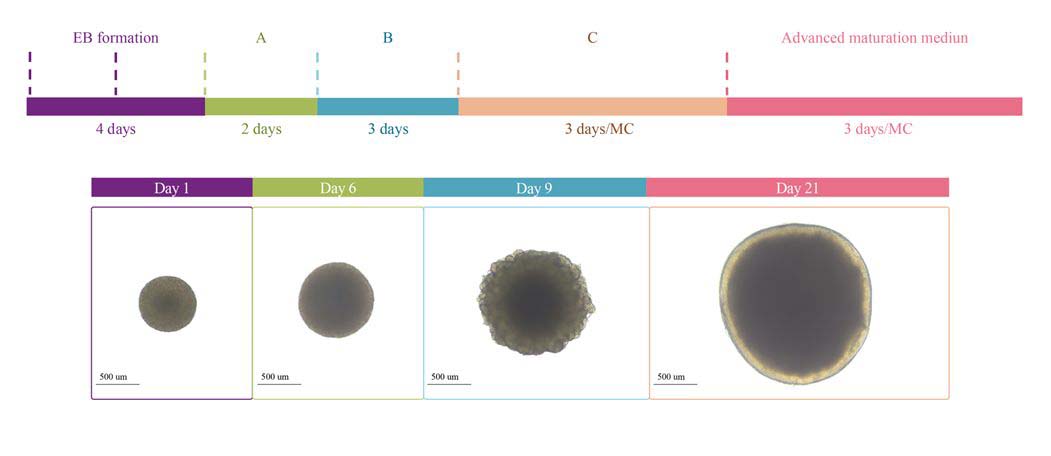
Protocol Diagram of cerebral organoid differentiation.
Orgnaoid Histology
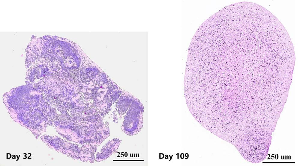
Left: Early-stage cerebral organoid show rosette-like structures (neural stem cells), which become smaller as organoids develop.
Right: Day 109 cerebral organoids show uniform morphology and show no dead core inside.
Marker Expression
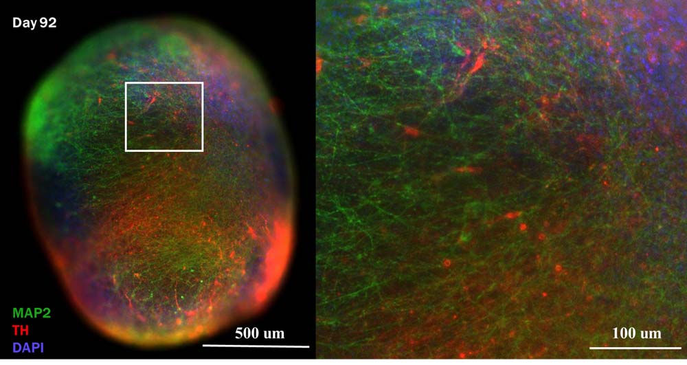
Presence of TH and MAP2 positive neurons in day 92 cerebral organoid.
TH: used as cell marker of dopaminergic neurons.
MAP2: mature neuron cell marker.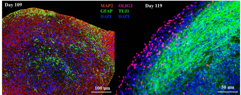
Left: Presence of GFAP positive cells at day 109 cultured cerebral organoid.
Right: Presence of OLIG2 positive cells at day 119 cultured cerebral organoid.
GFAP: marker for astrocyte.
OLIG2: marker for oligodendrocyte.
Organoid Activity
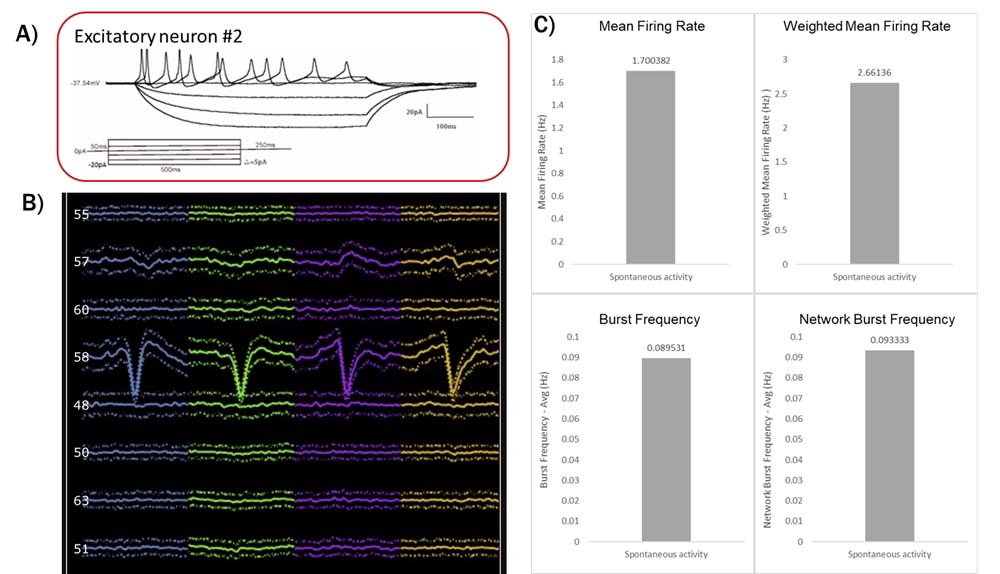
A: patch-clamp recording of excitatory neurons sectioned from cerebral organoids at day 102. Excitatory neuron showed stable response to step injection currents.
B: Recording of cerebral organoid (day 60) using silicon probe. Neurons showed spontaneous activities and different waveforms.
C: MEA recording indicated spontaneous bursting activities for cerebral organoid (day 86).
Organoid Application
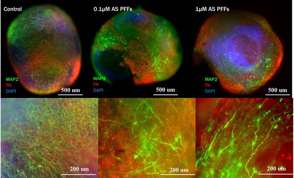
Using cerebral organoids for modelling Parkinson's Disease. Cerebral organoids are treated with Human Alpha-Synuclein Pre-formed Fibrils Protein. Dopaminergic neurons and MAP2 positive neurons are damaged with both 0.1 μM and 1 μM Alpha-Synuclein Pre-formed Fibrils.
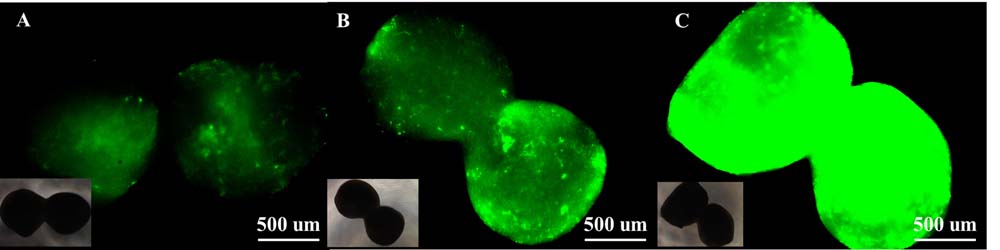
AAVs selection using cerebral organoids: Different capsid of AAVs were incubated with cerebral organoids. As the results, different affinities of each AAV to the cerebral organoid were observed.
用户评价 发表评论

 联系我们
联系我们 


 项目合作
项目合作