- English
- 日本語
- 한국어
- Deutsch
- Français
- Español
No data
官方服务号
招聘公众号

Die folgenden ELISA/SPR/BLI-Verfahrensbeschreibungen sind kostenlos erhältlich.

Immobilized Human Claudin-18.2 Full Length Protein-VLP (Cat. No. CL2-H52P7) at 5 μg/mL (100 μL/well) can bind Monoclonal Anti-Chimeric Claudin-18.2 Antibody, Human IgG1 with a linear range of 0.2-3 ng/mL (QC tested).
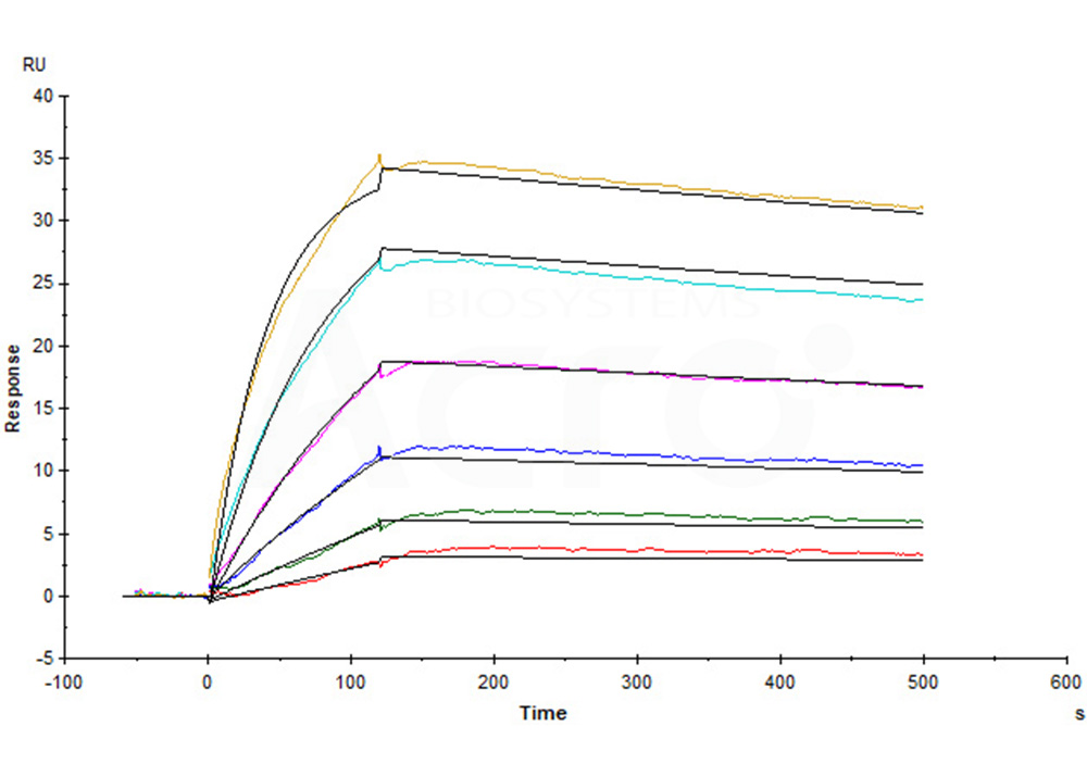
Human Claudin-18.2 Full Length Protein-VLP (Cat. No. CL2-H52P7) captured on CM5 Chip via Anti-Claudin-18.2 antibody can bind Anti-Claudin-18.2 antibody with an affinity constant of 0.374 nM as determined in a SPR assay (Biacore T200) (Routinely tested).
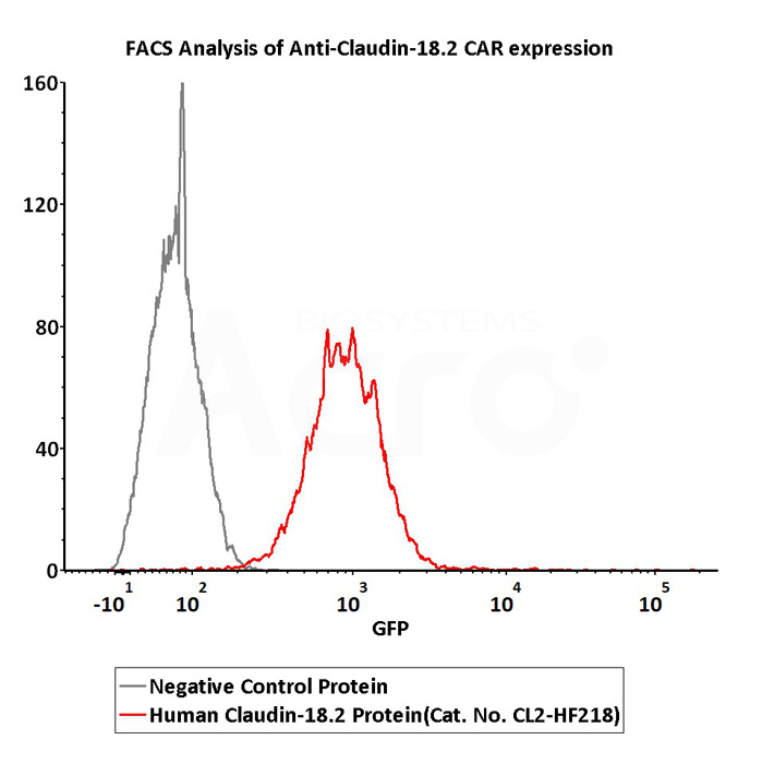
2e5 of Anti-Claudin-18.2 CAR-293 cells were stained with 100 μL of 3 μg/mL of Fluorescent Human Claudin-18.2 Full Length Protein-VLP (Cat. No.CL2-HF218) and negative control protein respectively, FITC signals was used to evaluate the binding activity (Routinely tested).

Immobilized Human Claudin-18.2, His,Twin-Strep Tag (Cat. No. CL2-H5587) at 2 μg/mL (100 μL/well) can bind Monoclonal Anti-Chimeric Claudin-18.2 Antibody, Human IgG1 with a linear range of 0.1-2 ng/mL (QC tested).
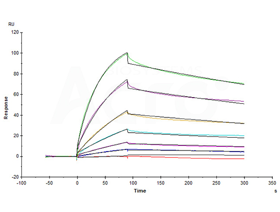
Anti-Claudin-18.2 mAb captured on CM5 chip via anti-Human IgG (Fc) antibody can bind Human Claudin-18.2, His,Twin-Strep Tag (Cat. No. CL2-H5587) with an affinity constant of 6.72 nM as determined in a SPR assay (in presence of DDM and CHS) (Biacore T200) (Routinely tested).

Immobilized Biotinylated Human Claudin-18.2 Protein, His,Avitag&Flag Tag (Cat. No. CL2-H85D4) at 1 μg/mL (100 μL/well) on streptavidin (Cat. No. STN-N5116) precoated (0.5 μg/well) plate can bind Monoclonal Anti-Chimeric Claudin-18.2 Antibody, Human IgG1 with a linear range of 0.1-2 ng/mL (QC tested).

Monoclonal Anti-Chimeric Claudin-18.2 Antibody, Human IgG1 captured on Protein A Chip can bind Human / Cynomolgus Claudin-18.2, His,Twin-Strep Tag (Cat. No. CL2-H5586) with an affinity constant of 12.6 nM as determined in SPR assay (Biacore 8K) (Routinely tested).
| Name | Research Code | Research Phase | Company | Indications | Clinical Trials |
|---|
 积分兑换
积分兑换 关注公众号
关注公众号
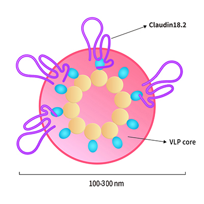
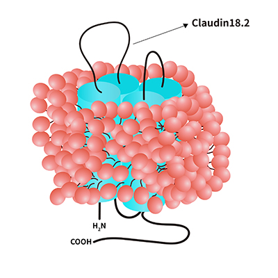
 Claudin 18.2-VLP